


Anatomical Models for the finishing touch
Visualizing 3D patient information from CT data can often reveal hidden facts. But having a tangible model of the anatomy in your hands adds another dimension to it. Physically reproducing your patient’s anatomy preoperatively allows you to come to a more precise diagnosis through the insight that it offers into the complexity of the specific pathology. Besides a better insight, you can also already practice your surgical approach; you can saw parts of the model, drill in it or prebend your plates on it. It enables you to go into surgery fully prepared, with an optimal treatment plan, optionally with a pre-bent plate, and avoid surprises. This reduces the time needed to perform the surgery, as well as increasing its accuracy and efficiency and ensures a better outcome for the patient.
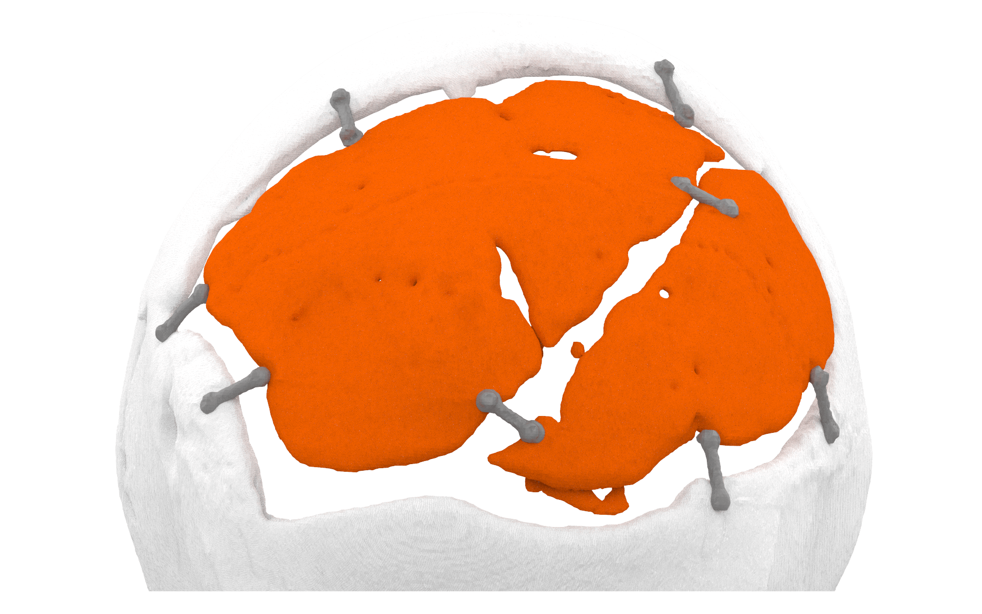
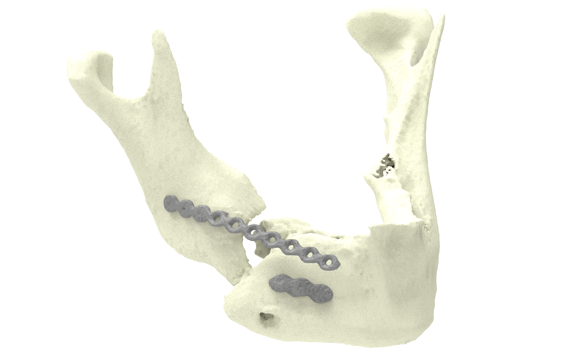
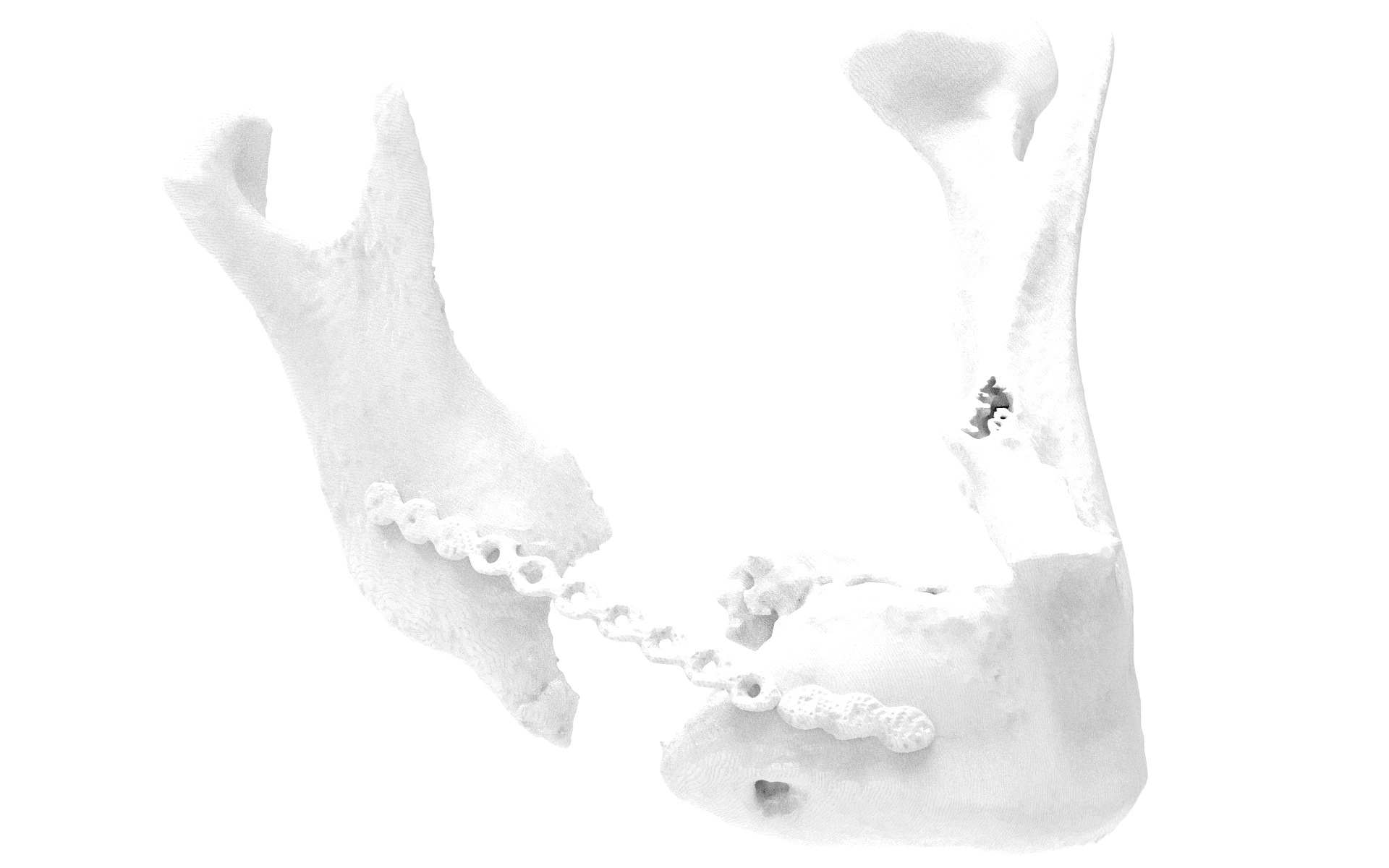
Fibula reconstruction of the mandible
Case by L. Poort, MD
A patient with bisphosphonate-related osteonecrosis of the mandible that developed a pathological fracture despite sequesterotomy of the right corpus mandibular. In order to bridge the defect, an osteosynthesis plate was inserted. This osteosynthesis plate later fractured and the patient was then referred to MUMC+. To bridge the bone defect, a fibula reconstruction of the mandible was planned. The jaw cups were repositioned in the correct position into the temporomandibular joint and the defect was filled with the fibula to reconstruct the patient’s original shape and position.
We employed the service of Xilloc to ensure optimal results. Using computer aided design, Xilloc repositioned the jaw cup into the temporomandibular joint and removed the fractured osteosynthesis plate. An anatomical model was then built using additive manufacturing and was subsequently utilized to bend a new osteosynthesis plate preoperatively to use during surgery. This enabled us during surgery to perfectly reposition the two mandibular parts and to accurately place the fibula to bridge the defect for an anatomical reconstruction.
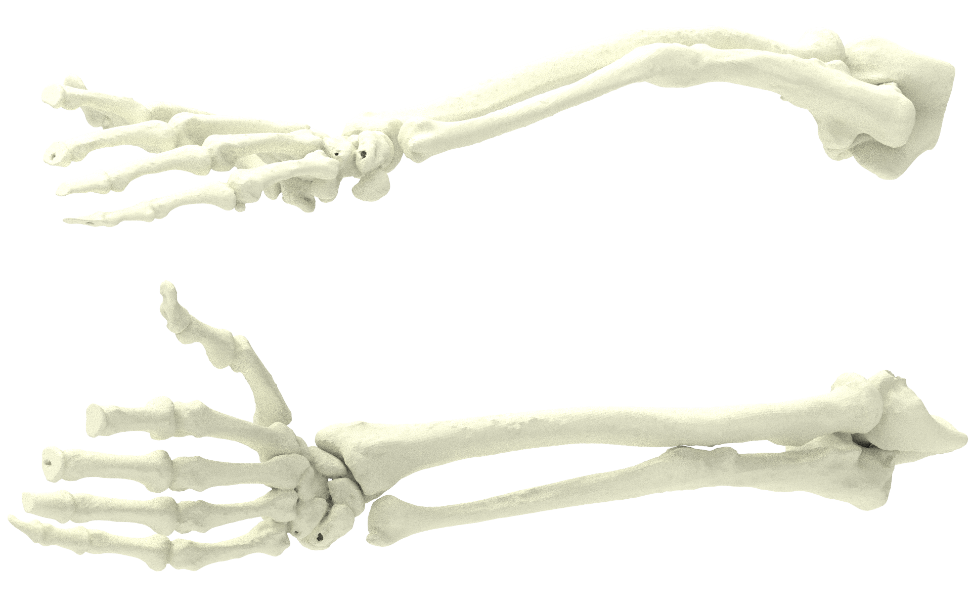
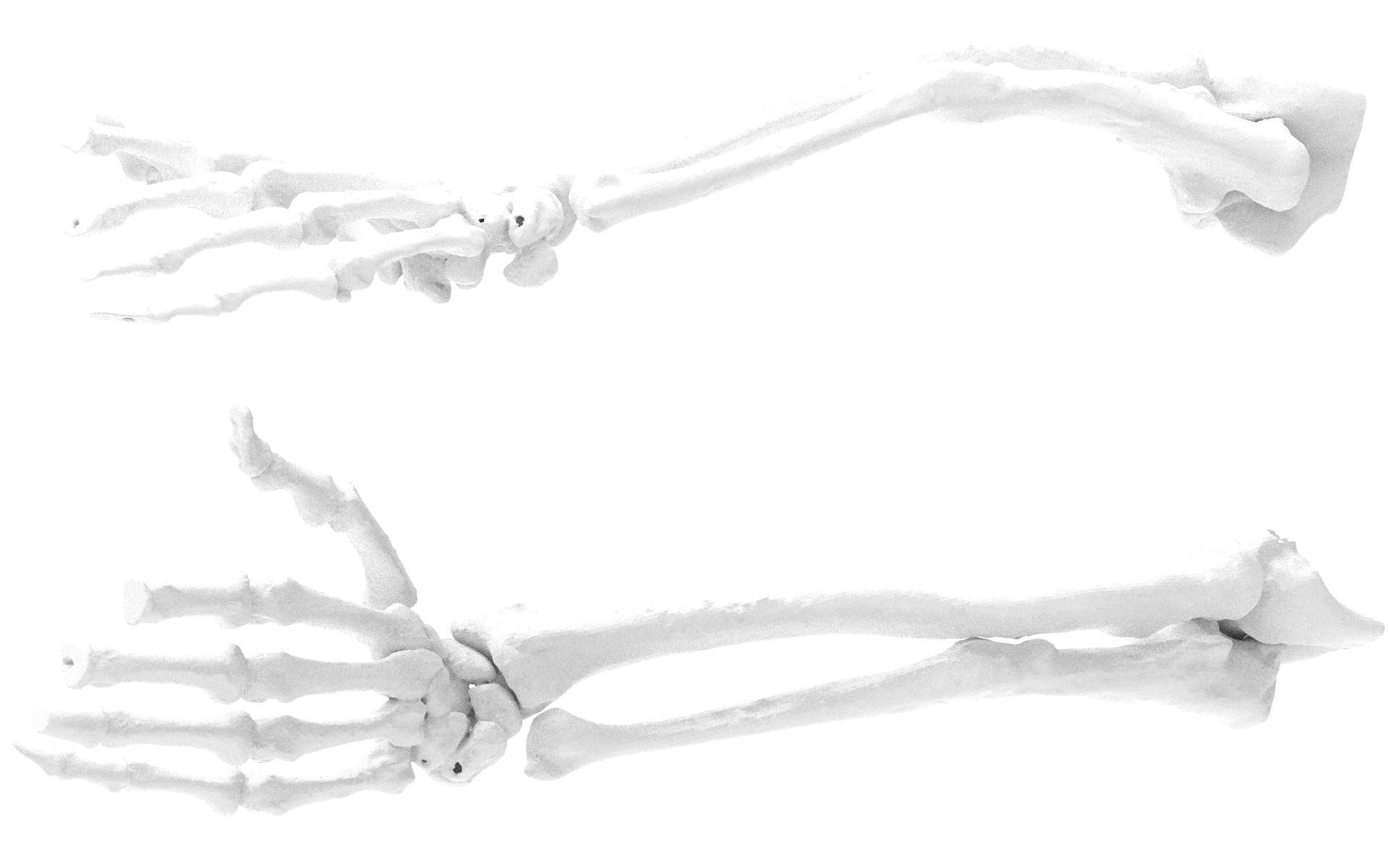
Antebrachii malunion
Case by G.A.M. Govaert, MD and Prof. Dr. P.R.G.Brink
When a patient has a complex malunion after an antebrachii fracture leading to rotational restriction, it can be difficult to identify the exact problem and decide how to best correct the situation. In particular, rotational errors require careful preoperative planning, and determining the appropriate osteotomy level is essential for a good outcome of the operation. The surrounding soft tissue is a crucial factor for the result. With a preoperative Anatomical Model from Xilloc and their medical engineering consultancy, we were able to reveal certain facts and help the patient by restoring normal antebrachii rotation.
Anatomical models to help repair a complex malunion