Surgical Guides to get it right

A virtual surgery plan is only valuable when you can translate it to the OR. An effective way to do so is by using surgical guides. A surgical guide is a small customized tool made from a sterilisable material that can be used for short-term in a patient and that guides your saw and/or drill in the planned direction. On request we can assist you with a full pre-operative surgical planning,  including the cutting planes and drill holes and their respective angles. To transfer this plan to the actual procedure, we design surgical (cutting) guides. The surgical guide fits exactly on a predetermined part of the patient’s bone, making sure you cut and drill at the right place, under the right angle (and to the right depth).
 
Guides can be used in any kind of surgery (osteotomies), but are often used for mandible reconstruction with an autologous bone graft (fibula or iliac crest) or for meningioma resection. To illustrate we present here a mandible resection and reconstruction case using an iliac crest graft and below a similar mandible reconstruction with a fibula graft.

Case 1: Mandible resection and reconstruction with iliac crest

Segmental resection of the mandible followed by (free flap) mandible reconstruction with autologous bone is a delicate procedure that can benefit from careful 3D surgical planning. From a CT-scan, accurate 3D bone models can be generated, which can be used in surgical planning software. You don’t need to invest money to buy, or time to learn this software, but our biomedical engineers can do this with you. Together we define the cutting planes and make a virtual resection of the mandible. Then we find the optimal position of the iliac crest within the resected mandible to achieve a good mandible reconstruction. To accurately transfer the planning to the operating room, customized surgical cutting guides are then designed by our engineers and 3D printed. These cutting guides ensure that the resection of the mandible and the resection of the bone from the donor site (here the iliac crest) are executed precisely according to plan.

Two surgical cutting guides are designed and 3D printed in polyamide. These cutting guides fit perfectly on the mandible and the pelvis in only one orientation and indicate the outline of the planned resection area. Based on the surgeons preference, the cutting guides are either equipped with slots to cut through (see the mandible guide on the left) or with flanges to cut along (see the pelvis cutting guide on the right). The guides can be fixated onto the bone with several screws.


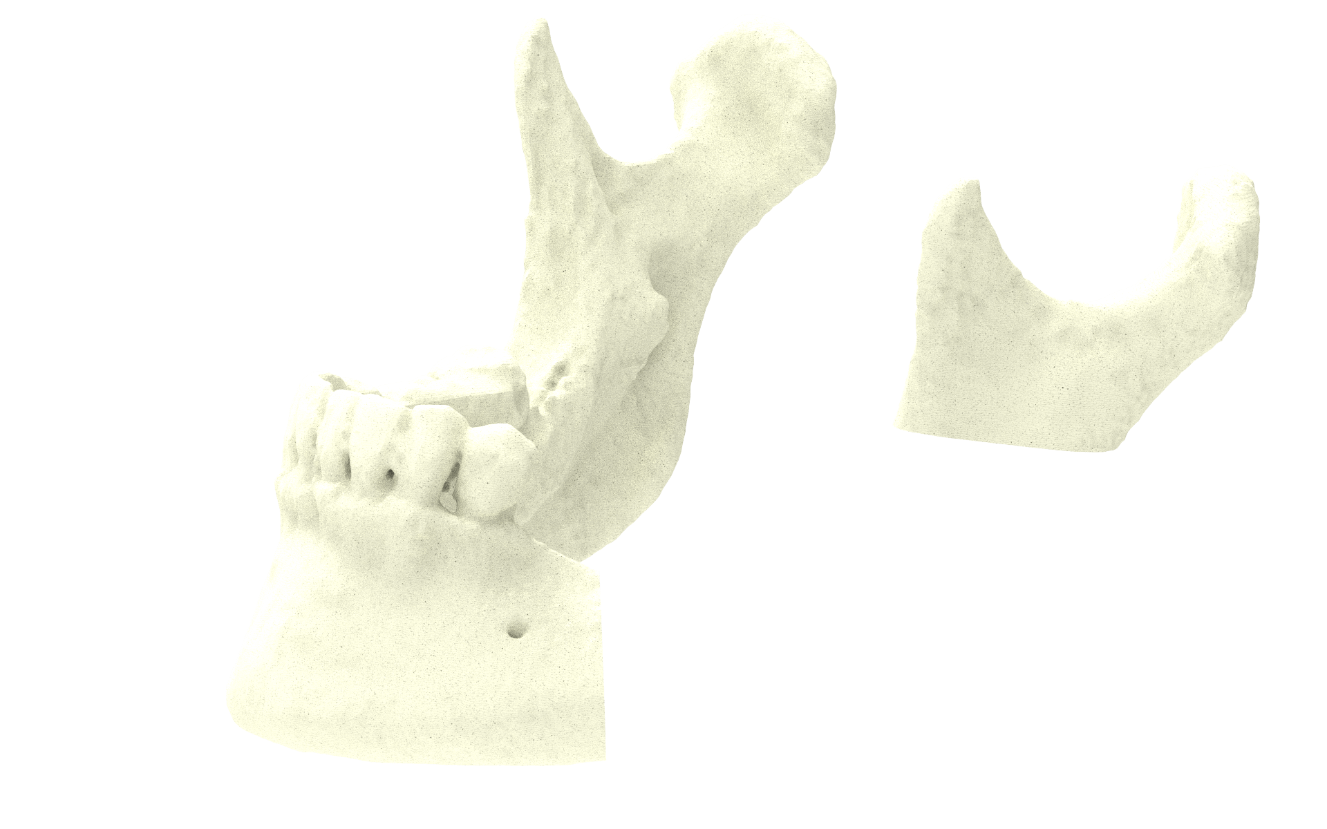
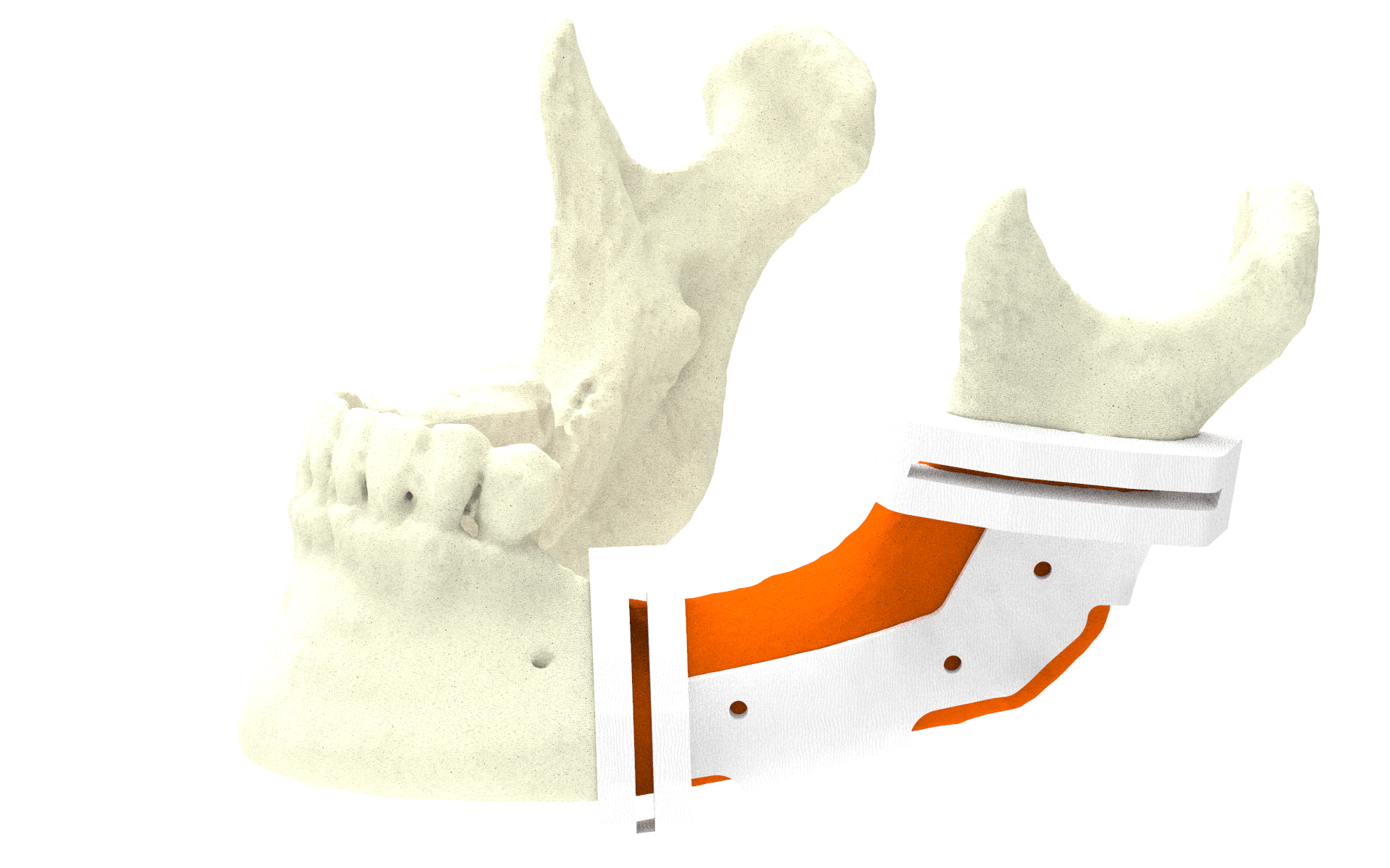
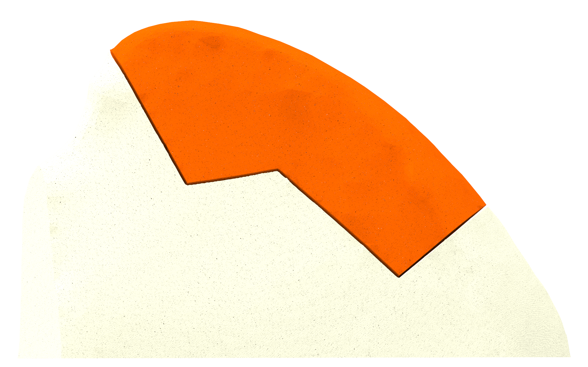
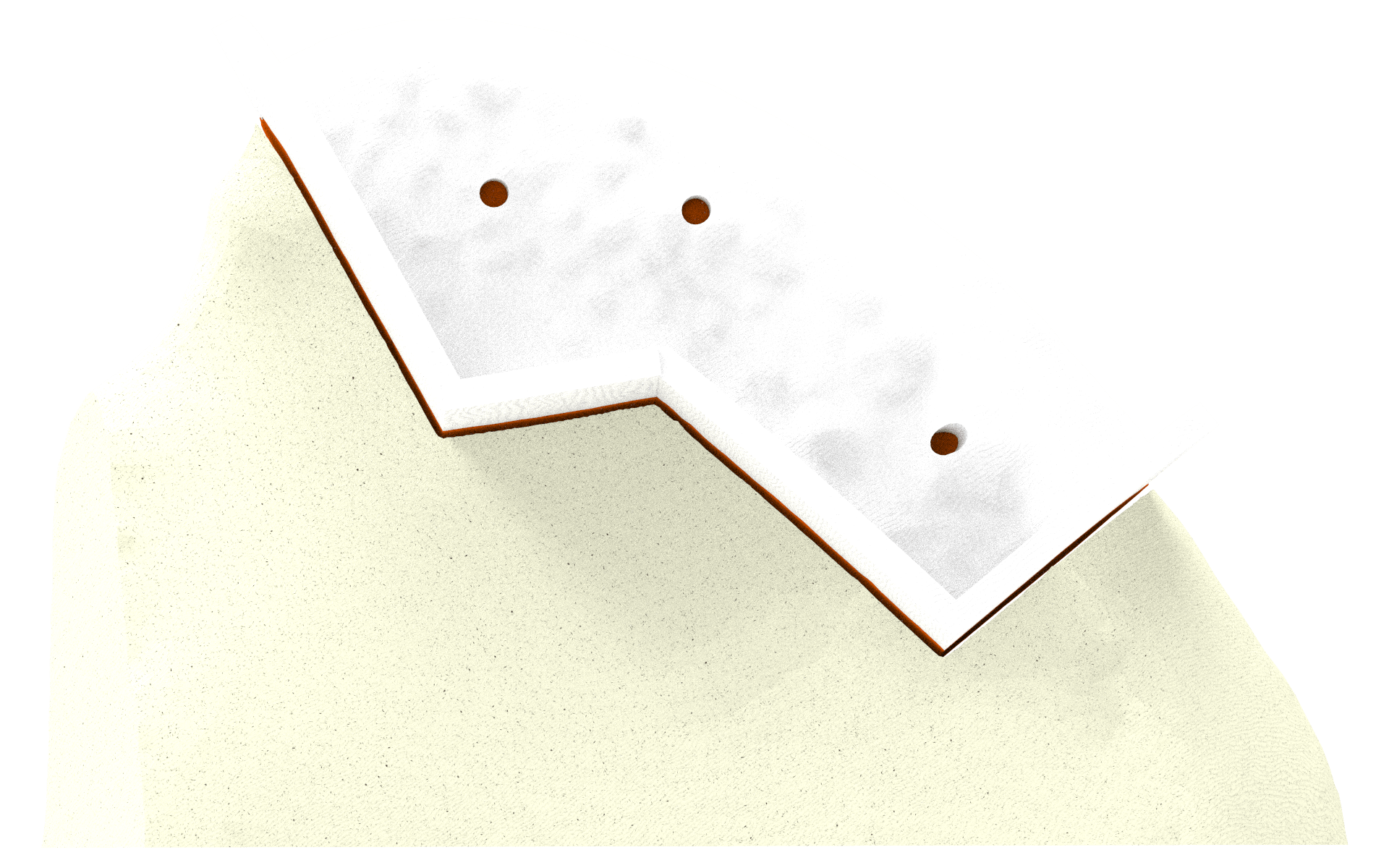
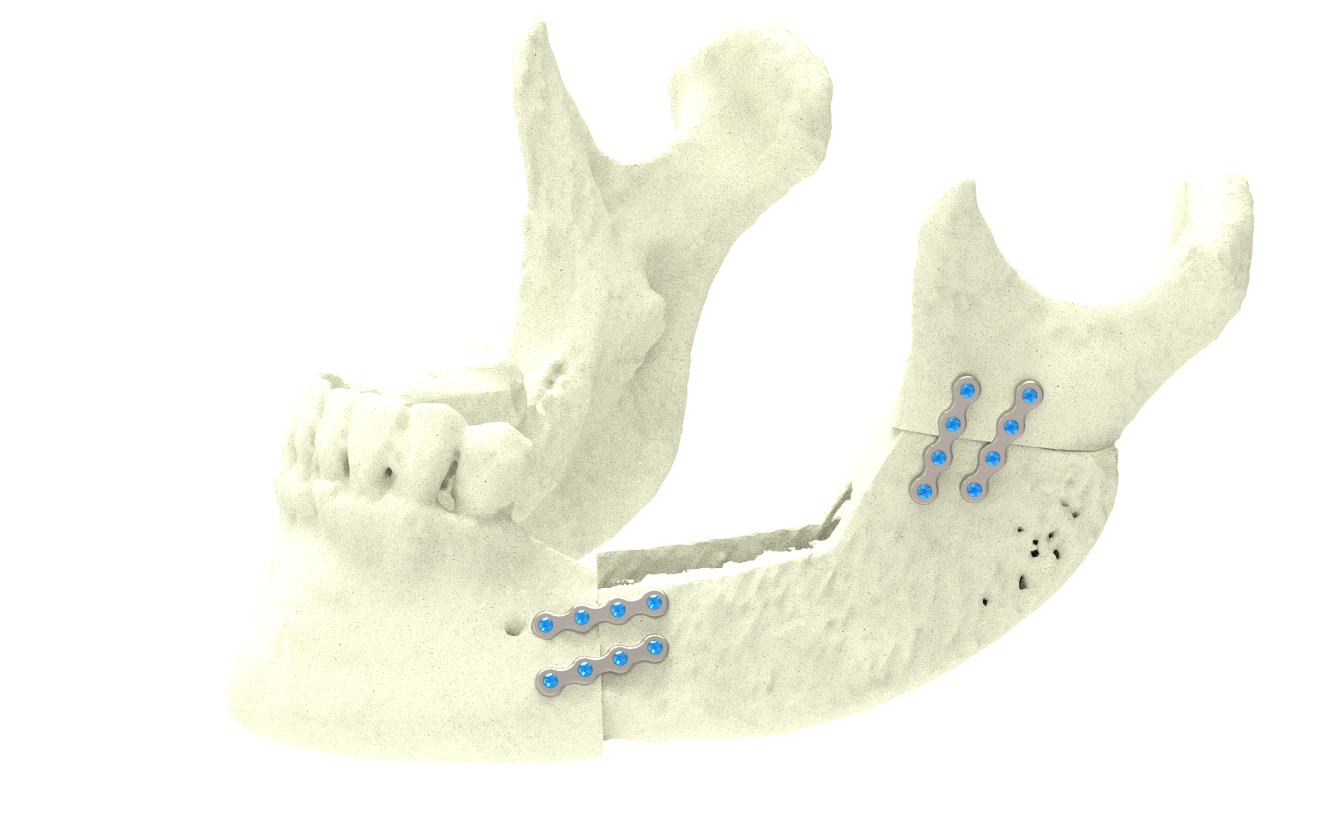
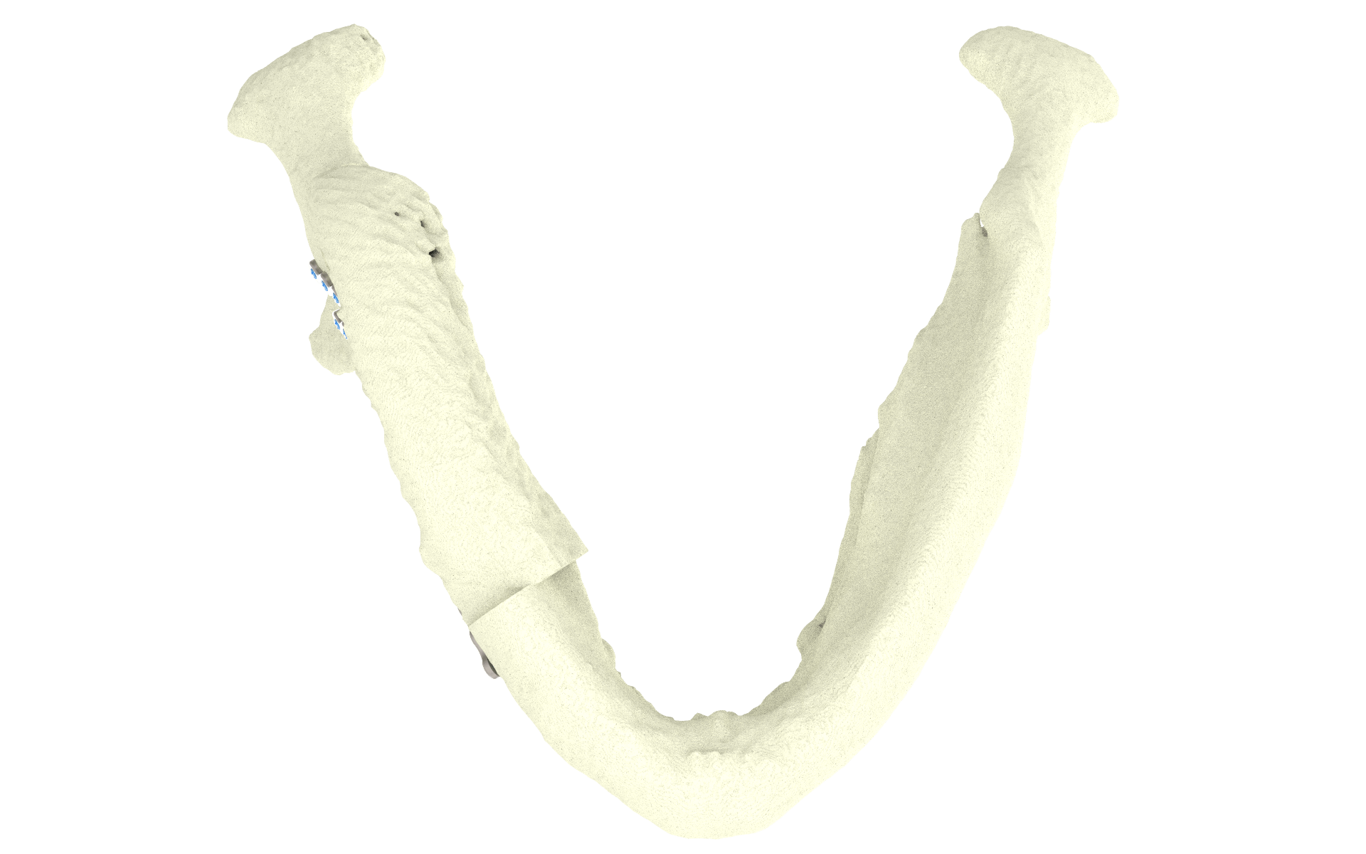
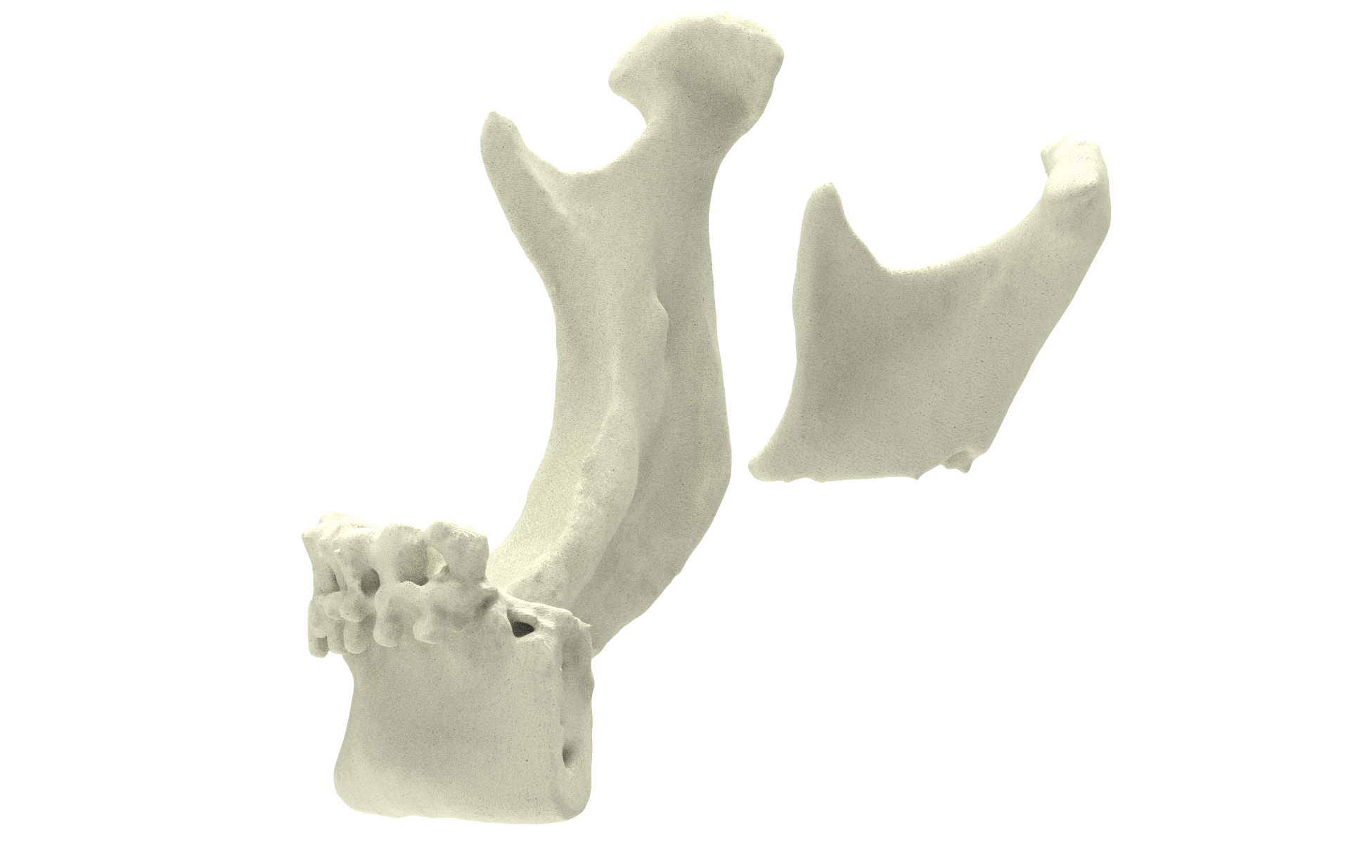
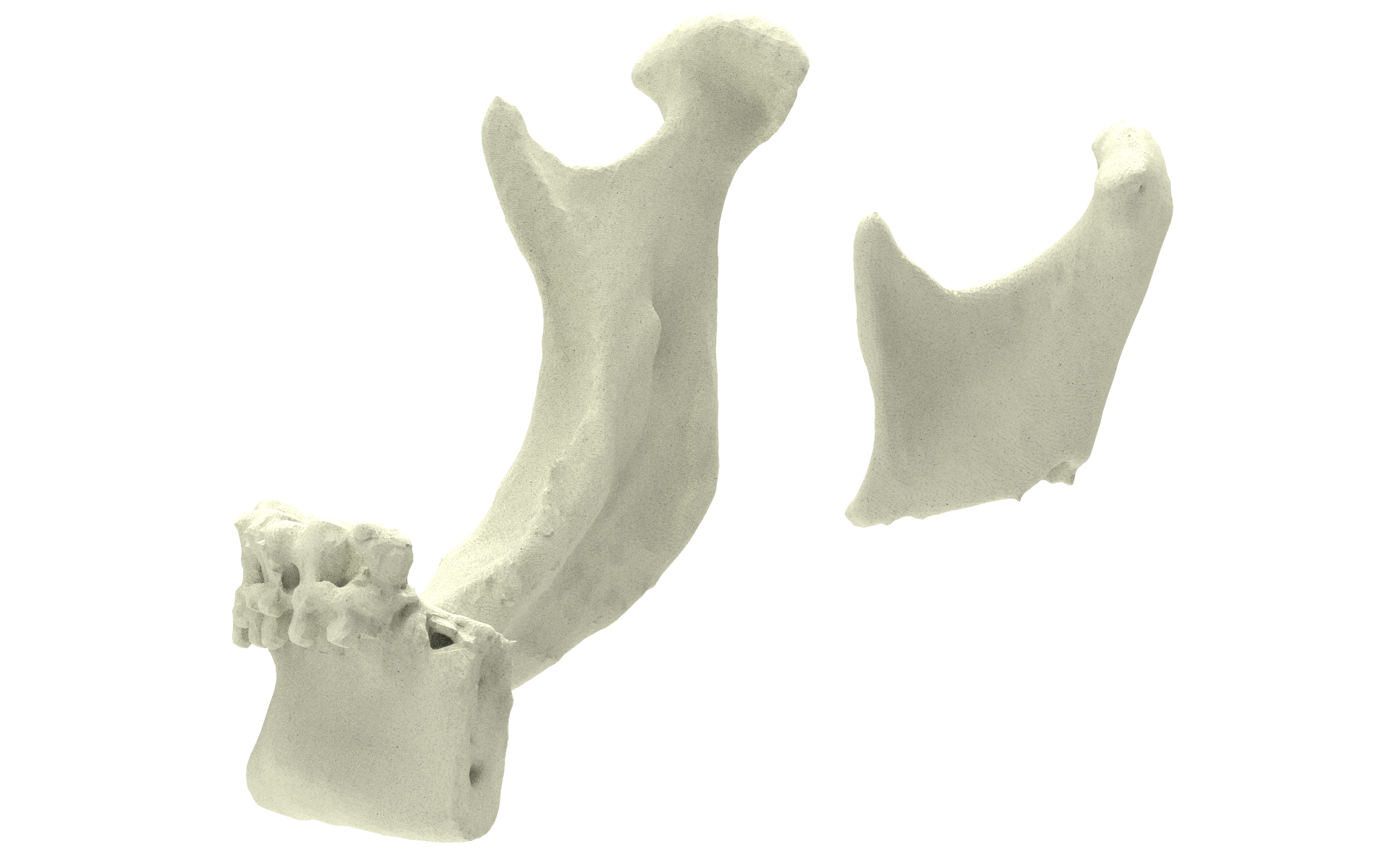
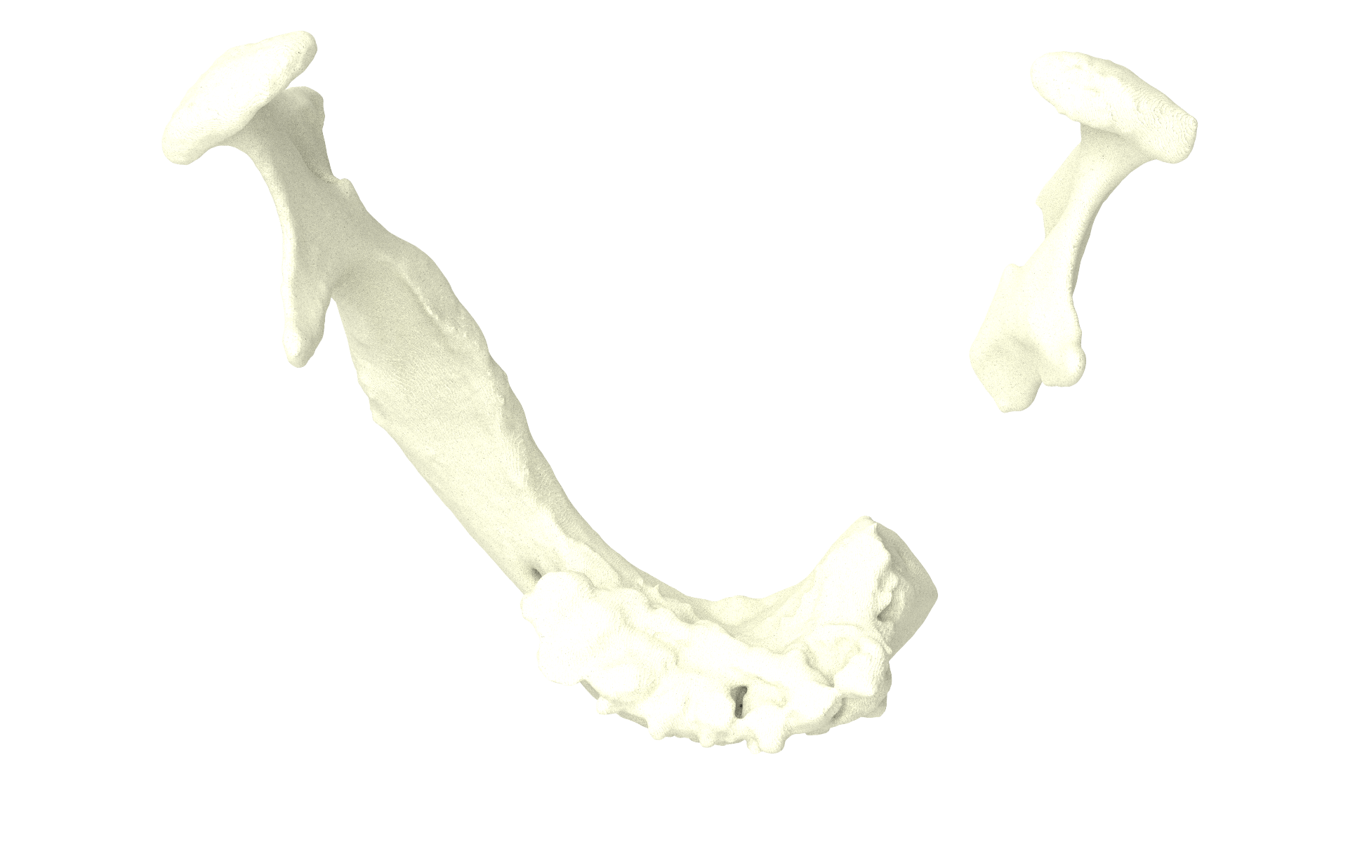
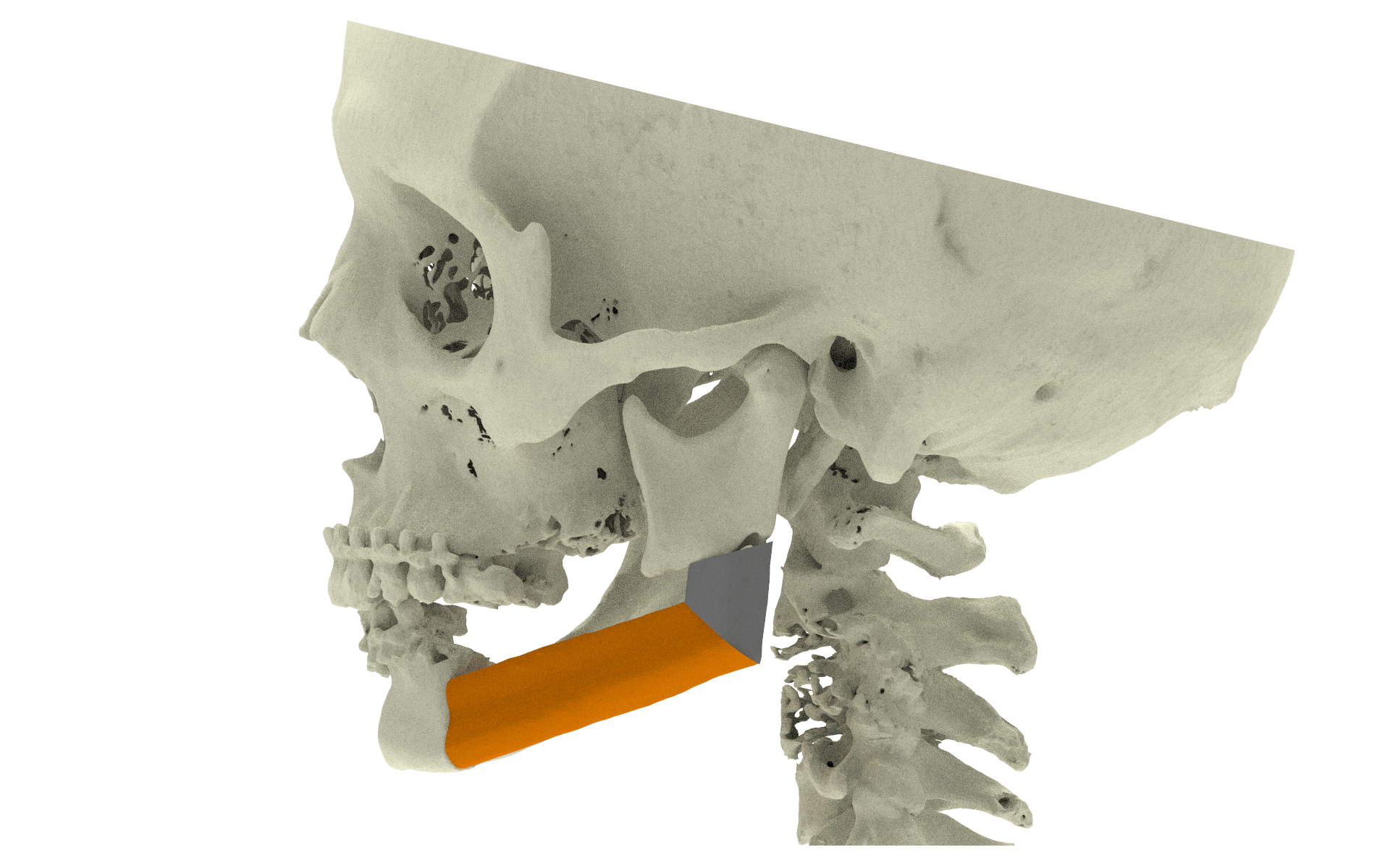
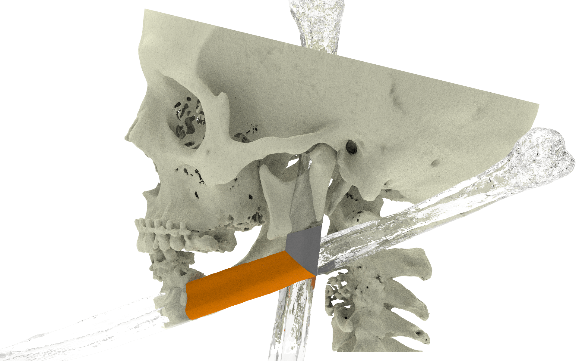
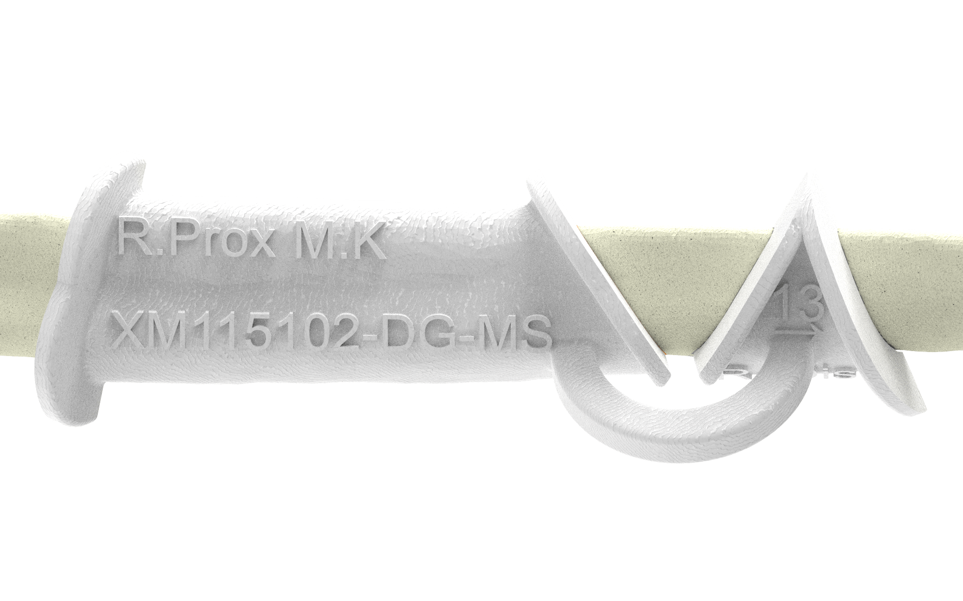
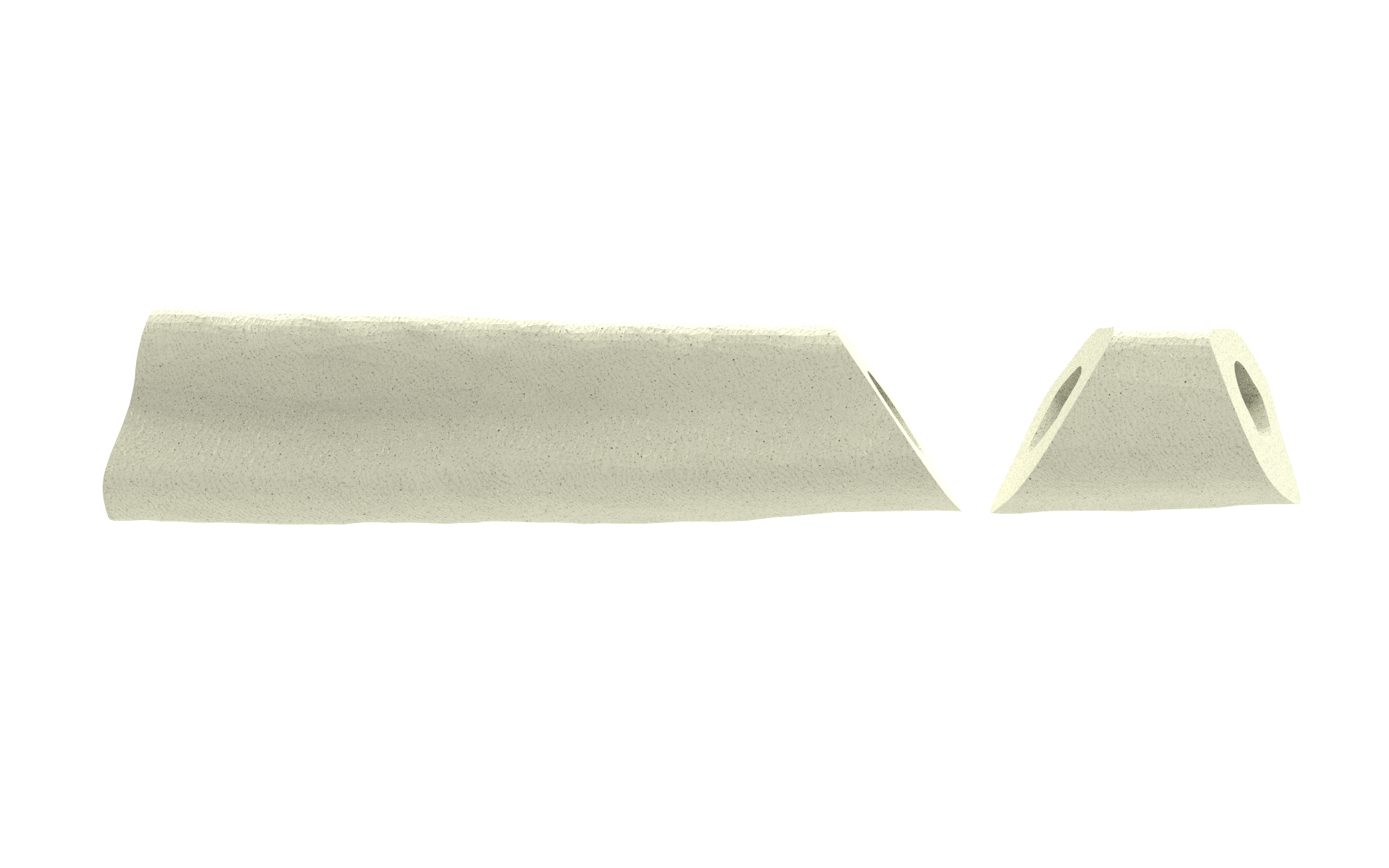
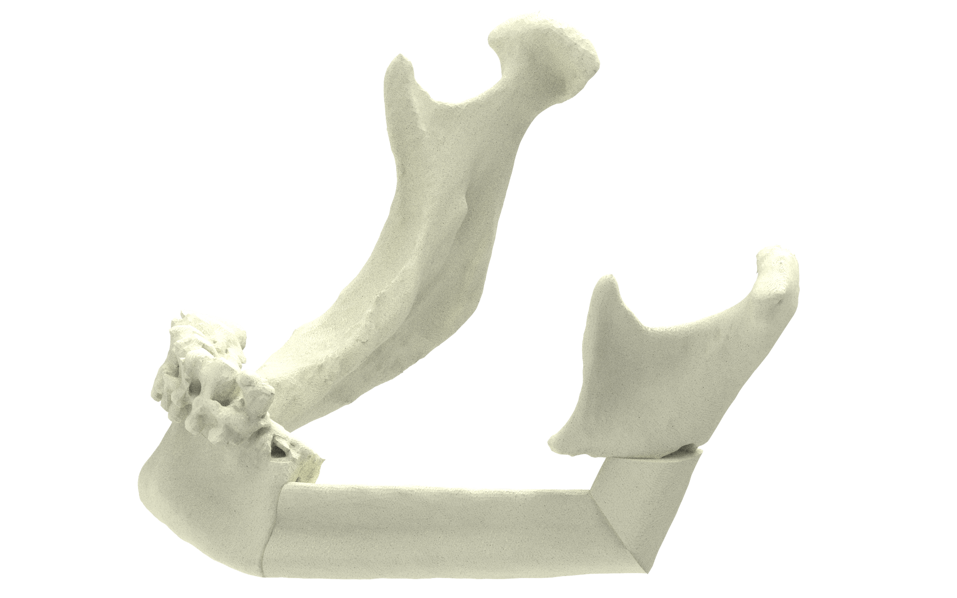
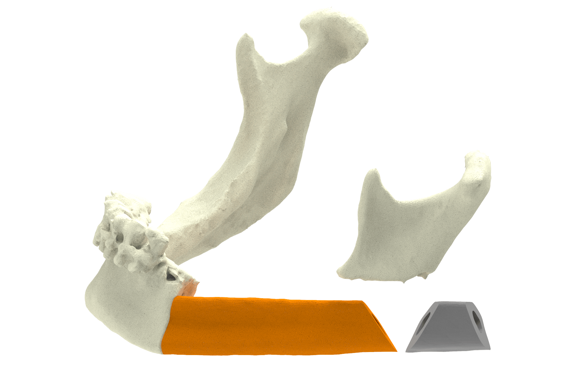
When you approve the planning and design, the cutting guides are produced by a professional 3D printer (Selective Laser Sintering) according to our protocol for medical production (conform ISO 13485). Afterwards they are thoroughly cleaned, packed and shipped. The guides will be delivered to the hospital on the day requested by the surgeon, but the latest one day before surgery, so they can be sterilized. The polyamide cutting guides can easily be sterilized using an autoclave and sterilization guidelines are provided. Optionally, an accurate anatomical model of the resected mandible can be provided for prebending of the osteosynthesis plates.


During surgery the bone is exposed and the cutting guides will fit perfectly on the mandible and iliac crest, allowing resection of the mandible according to plan. The resected part of the iliac crest can be positioned in the defect of the mandible and deep circumflex iliac artery and vein can be connected to vessels in the neck to provide blood supply. Then the (prebent or standard) osteosynthesis plate(s) can be used to fixate all the parts together.


The surgeon was very pleased with the result as seen on the postoperative CT scan. The esthetic outcome made the patient smile again.

Case 2: Mandible reconstruction with fibula


Backward planning

A pre-operative virtual planning and the associated surgical guide(s) will assist you to drill and saw in the right direction, according to plan.
“Backward” planning can be used to find the best position of the bony parts. You start by predefining the optimal position of the mandibula, which determines the void that needs to be bridged by the bone graft.

Dislocated mandibula

Virtual repositioning

Optimal mandibula position

Matching the fibula to “bridge the gap”

Using a Computed Tomography (CT) scan of one of the legs, the necessary bony shape can be virtually matched from the patient’s fibula.

Necessary fibula segments


The cutting guide

The guide is designed in such way that it matches the patient’s anatomy perfectly and only fits on the region determined above. For the surgeon to know where to open the patient’s leg, we can indicate the approximate position, in centimeters distance from the ankle point. This is done by engraving a number in the guide, number 13 in the example below.

Cutting

Cutting along the four angular planes will create the two pieces of fibula needed for the mandibula reconstruction.

Graft

Now you have two perfect graft pieces, ready to be placed.

Placing

The fibula pieces match the jaw and each other.

Fixating

Together, they make the right angle.